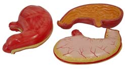
XC-306 Stomach Model |
This model shows the morphology of the stomach
in a moderate distended state. With the longitudinal
section, the model shows the structures of the
gastric folds, pyloric valve, pyloric sphincter
muscles, gastric mucosa and the transitional
mucosa of the gastricesophagus. Made of hard
plastic and magnified 2 times the natural size. |
Click to Enlarge |
|
 |
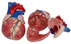
XC-307 Jumbo Heart Model |
This model helps the students to understand the
external features and internal structures of the
heart, and its relation with the large blood vessels.
Thus a clearer conception of the routes of the
systemic and pulmonary circulation can be
obtained. Dissectible into 3 parts, 4 times enlarged.
Made of PVC plastic. |
Click to Enlarge |
|
 |
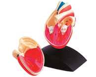
PD-002 Heart Model |
- Full size heart model
- Durable ABS Base
- Detachable Cover
- Highly Realistic Decoration
|
Click to Enlarge |
|
 |
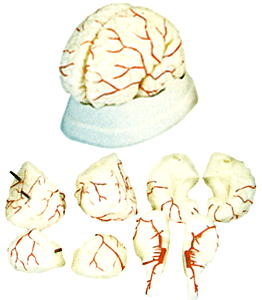
XC-308 Brain With Arteries |
This model facilitates the students to get a correct
understanding of the external features of the brain and its
arterial supply as a whole, as well as the relations between
their component portions. External features of the brain :
cerebral hemisphere, brain stem, cerebellum. The arterial
supply of the brain : sources, vertebral, internal carotid
arteries, arterial supply of the cerebellum and cerebrum.
|
Click to Enlarge |
|
 |
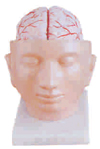
XC-318 Brain With Arteries on Head |
This model shows the brain structure
inside the skull. Two halves brains can be
disassembled into: frontal with parietal
lobes, temporal with occipital lobes, half
of brain stem, half of cerebellum.
Life-size, dissectible into 9 parts. |
Click to Enlarge |
|
 |
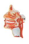
XC-324 Model of the Head |
This model shows the
interior parts of the mouth
cavity and pharynx with
network of vessels and
trigeminal nerves.
|
Click to Enlarge |
|
 |
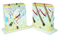
XC-313 Enlarged Skin Model |
A greatly enlarged (105 times) cross sectional view of the human skin
showing three layers and a close-up view of a hair follicle, sweat gland,
fatty tissue and more. Front side and back view. Not dissectible. Shows
the structures of the human scalp as follows:-
- Structure of the skin : epidermis, dermis, hypodermis
- Appendages of the skin : the sweat glands, the sebaceous glands,
the hairs
- Blood vessels and nerves of the skin
- Mounted on a plastic base.
|
Click to Enlarge |
|
 |
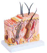
XC-313-2 Skin Block Model |
This model shows a section of human skin in three dimensional forms.
Hair, sebaceous and sweat glands, receptors, nerves and vessels are
shown in detail. 70 time enlarged. |
Click to Enlarge |
|
 |
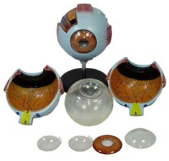
XC-316 Giant Eye Model |
The different parts of the eyeball model are detachable to show the
following structures:
- Tunica External : Showing cornea and sclera with attachments
of ocular muscles and optic nerves
- Tunica Media : Showing the iris, the ciliary body and the
chorioid
- Tunica Internal is Retina
- Refraction Media : Showing the lens and the vitreous body.
The model is made of PVC. |
Click to Enlarge |
|
 |
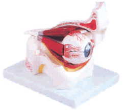
XC-316B Eye with Orbit |
|
The model shows the eyeball with optic nerves and muscles in
its natural position in the bony orbit. Dissectible into 10 parts.
3 times enlarged. |
Click to Enlarge |
|
 |
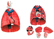
XC-320 Larynx, Heart and Lungs Model |
A life-size model separated into 7 parts. The lungs have 2
removable lobes to show the internal structures, the heart
bisects showing atria, ventricles and valves, the larynx
bisects and the diaphragm is shown. |
Click to Enlarge |
|
 |
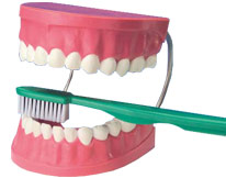
TH-001 Giant Dental Care Model with Toothbrush |
- 2.0x full size model
- Giant teeth
- Giant toothbrush
|
Click to Enlarge |
|
 |
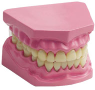
TH-002 Dental Model |
- 1.5x model with Hinge
- Realistic Shape and Details
|
Click to Enlarge |
|
 |
XC-309 Model of the Anatomical Nasal Cavity |
This model helps the students to understand the external and internal structures of the
nasal cavity.
- External nose : shows the section of the nasal bones and cartilages.
- Nasal cavity : on the lateral nasal wall shows the superior, middle and
inferior nasal chonchae project medially into the nasal cavity
forming the superior, middle and inferior nasal maxillary
sinuses.
- Paranasal sinuses : shows the frontal, sphenoid and maxillary sinuses.
Magnified ½ the natural size. Made of PVC.
|

Click to Enlarge |
|
 |
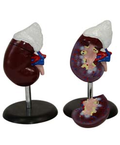
XC-310-3 Human Kidney With Adrenal Gland |
This detailed life-size model features the kidney, adrenal gland, renal and adrenal vessels
and upper portion of the ureter. Dissectible into 2 parts to reveal the cortex medulla, cortex
vessels and renal pelvis. Model can be removed from the stand for instruction and patient
education. |
Click to Enlarge |
|
 |
XC-312 Liver Model |
An economical way to study the basic structure of the liver. The complex vessels network
in the opened liver, displayed in different colors on this model : hilus vessels, extrahepatic
and intra-hepatic bile ducts. Liver mounted on stand. |
Click to Enlarge |
|
 |
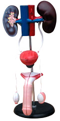
XC-331 Male Urogenital System |
This is intended for middle schools as a visual aid in teaching physiology
and hygiene to help the students to understand the external features of
the urogenital system and the internal structures of the kidney, urinary
bladder, penis and testicle. Made of soft PVC plastic. |
Click to Enlarge |
|
 |
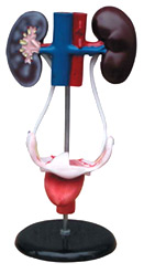
XC-332 Female Urogenital System |
This model serves as a model for teachers to teach students the female
urogenital system in schools and colleges. The model shows kidney,
ureters, urinary bladder, uterus, accessories of uterus, vagina, ovary
membrane, ligaments, uterus ligament and its artery etc. Made of PVC
plastic. |
Click to Enlarge |
|
 |
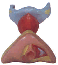
XC-429 Female Internal & External Genital Organs |
This model is served as a visual aid in teaching human anatomy in
nursing schools and medical colleges. It shows the entire female genital
organs including uterus, uterus appendix (fallopian tube, ovary and its
membrane, ligament teres uteri, ligament ovary proprium), vagina and
the structure of external genital organ. Part of uterus and vagina are
dissected to show its internal structures. Size: life-size. |
Click to Enlarge |
|
 |
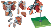
GD/A15102 Male Genital Organs |
It is made of advanced PVC material.
The model is dissected in median and mounted
on a stand. It shows the penis, prostate, bladder,
seminal vesicle, spermatic cord and inguinal
canal and base. 72 positions displayed. |
Click to Enlarge |
|
 |
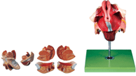
GD/A15105 Female Genital Organs |
It is made of Advanced PVC material.
The model consists of 4 parts and is mounted on
a stand. It shows the internal and external
female genital organs. 40 positions displayed. |
Click to Enlarge |
|
 |
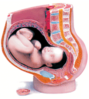
XC-332B Human Female Pelvis Section (4 Parts) |
This model is a median section, showing female genital organs with
bladder and rectum. The abdominal and pelvis muscles are shown
detailed.
Made of PVC plastic.
|
Click to Enlarge |
|
 |
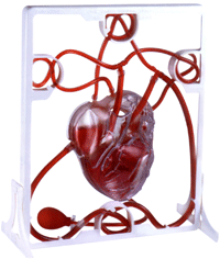
MK-019 Pumping Heart |
- A 3-D working plastic heart model with clear chambers. Squeeze bulb pumps simulate blood through clear heart chambers just like
real one.
- Complete heart model scale with display stand.
- Complete instruction manual, easy to assemble.
- Acrylic Paints and Brush are included.
|
Click to Enlarge |
|
 |
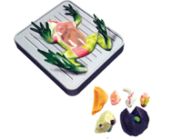
VT021 Frog Dissection Kit |
- Authentically detailed frog Body
- Removable soft texture organs
- Cross sections show muscle structure
- Glow-In-Dark "X-RAY" included
- Child safe lab tools included
|
Click to Enlarge |
|
 |
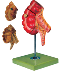
GD/A 12006 Appendix and Caecum |
Natural size and mounted on a stand. The model shows wall of the caecum and appendix. Separated into 2 parts. 17 positions are displayed.
Material: Advanced PVC and painted with imported paint. |
Click to Enlarge |
|
 |
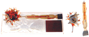
GD/A 18001 Neuron |
This model consists of 2 parts as: neuronal body and nerve fibre. It shows neuronal axon hillock, dendritic arbor, neurofilament, etc. 15 positions are displayed.
Material: Advanced PVC and painted with imported paint. |
Click to Enlarge |
|
 |
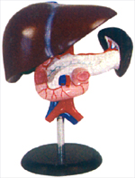
XC-311 Liver, Pancreas and Duodenum Model |
An economical way to study the basic structure of the liver, spleen, blood
vessels and pancreas. External structure is illustrated as well as the
pancreatic duct of the pancreas. This model also shows the abdominal
aorta and inferior vena cava. Life size. Dissectible into 3 parts.
Made of PVC. |
Click to Enlarge |
|
 |
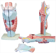
XC-301 Magnified Human Larynx Model
|
A functional model that demonstrates movements of the epiglottis and cartilages in
the voice box. It helps the students to understand the morphology and structure of
the respiratory tract and phonetic organ. On base 3 times enlarged. 3 parts
dissectible. |
Click to Enlarge |
|
 |
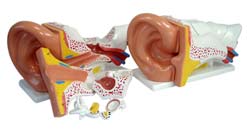
XC-303C New Style Giant Ear Model |
A giant model of the Ear, for elementary science classes,
shows the three main structural parts of the hearing organ
(external ear, middle ear, internal ear) and the position of the
equilibrium organ of human body.
External Ear : showing the shape of the auricle and the
primary features of the external auditory meatus.
Middle Ear : showing the type of membrane, the three
auditory ossicles (hammer, anvil, stirrup) and the
eustachian tube.
Internal Ear : showing the vestibule, cochlea and the three
semicircular canals of the osseous labyrinth.
|
Click to Enlarge |
|
|
 |
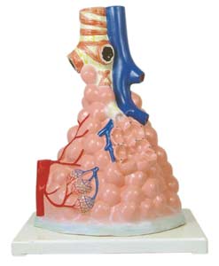
XC-302 Magnified Human Pulmonary Alveoli Model |
This model shows the small branches of principal bronchus:
- Section of bronchiole of no cartilage
- The relation between pulmonary alveoli and terminal bronchiole
- The structure of alveolar sac and alveolar duct
- The capillary rete in the alveolar sapta
Made of PVC plastic and mounted on plastic base. |
Click to Enlarge |
|
 |
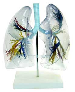
XC-330 Model Of the Transparent Lung Segment |
This model presents 10 segments in the right lung and 8 in the left. The
distributions of the bronchial tree can be observed through the transparent
lungs.
- Right lung: right upper lobe (3 segments), right middle lobe (2 segments), right inferior lobe (5 segments)
- Left lung: left upper lobe (4 segments), left inferior lobe (4 segments)
- Distribution of the bronchial tree: right bronchus, left bronchus.
- Hilus of lung.
This lung is made of transparent plastic. The trachea and bronchial tree is made of PVC. |
Click to Enlarge |
|
 |
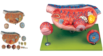
GD/A41003 Ovary Model |
This model consists of 8 parts, including ovary, primary
ovarian follicles, secondary ovarian follicles, ovium,
ovulation, ovum, corpus luteum, corpus albicans. It
demonstrates the procedure of ovum's development. 10
positions are displayed.
Material: Advanced PVC and painted with imported paint. |
Click to Enlarge |
|
 |
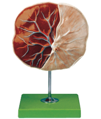
GD/A42010/1 Human Placenta |
The model shows the structure of placenta and the relation
between placenta and umbilical cord.
7 positions are displayed.
Material: Advanced PVC and painted with imported paint. |
Click to Enlarge |
|
 |
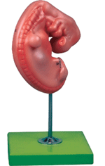
GD/A42004 Embryo |
The embryo model is of about 4 weeks old and mounted on a stand. 12 positions are displayed.
Materials: Advacned PVC and painted with imported paint. |
Click to Enlarge |
|
 |
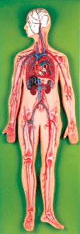
GD/A16001 Circulatory System |
This model shows general network of the vessels body and it displays around 81 positions. It is made of advanced PVC materials. |
Click to Enlarge |
|
 |
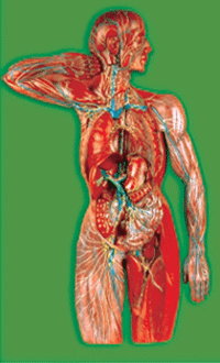
GD/A16011 Lymphatic System |
This model shows the component and structure of the lymphatic system and it displays around 63 positions. It is made by Advanced PVC materials. |
Click to Enlarge |
|
 |
GD/A18101 Nervous System |
This model shows the structure of central nerves, peripheral nerves
including spinal nerves radiated from central systems to other parts of
body (such as ulnar nerve, radial nerves of brachial plexus, median
nerve, ischiadic nerve) and it displays around 33 positions. It is made by Advanced PVC materials. |

Click to Enlarge |
|
 |
GD/A18102 Spinal Cord in the Spinal Canal |
This model shows ventral side, brainstem and spinal cord. Branches
up to the coccygeal plexus, sympathetic trunk with its connections to
the central nervous system and it displays around 47 positions. It is made by Advanced PVC materials. |

Click to Enlarge |
|
 |
GD/A18110 Sympathetic Nervous System |
This model shows the outlook of automatic nervous system, including
sympathetic nervous system and parasympathetic nervous system.
The sympathetic one shows in yellow color and parasympathetic one in
red; and it displays around 53 positions. It is made by Advanced PVC materials. |

Click to Enlarge |
|
 |
XC-315 Digestive System |
Life-size digestive system model,
demonstrates the entire digestive
system in graphic relief. Digestive
system features : nose, mouth
cavity and pharynx, esophagus,
GI tract, liver with gall bladder,
pancreas and spleen.
The duodenum, caecum and
rectum of the digestive system
are opened. Dissectible into 3
parts. Mounted on baseboard.
|

Click to Enlarge |
|
 |


